

Quick answer: Tuberculosis can affect the brain as tuberculous meningitis or as tuberculomas. It commonly causes hydrocephalus, because inflammation blocks the flow and absorption of cerebrospinal fluid. Early symptoms are vague — weeks of fever, headache and weight loss — which often delays diagnosis.
Brain TB and Hydrocephalus: Vital Facts Every Family Must Know
By Haris Bin Tahir
Father of a hydrocephalus survivor. Independent researcher. Not a doctor.
Founder, Brain Care Path · braincarepath.com
Key Takeaways
- Tuberculous meningitis — TB infection of the brain’s lining — is one of the leading infectious causes of hydrocephalus worldwide, particularly in children under five
- The connection between TB and hydrocephalus is frequently missed or delayed because initial TB symptoms can appear in the lungs first, masking what is happening in the brain
- Early recognition of this specific pathway — infection leading to brain inflammation leading to hydrocephalus — changes how quickly families receive the right treatment
The first diagnosis was lungs.
TB in the lungs. We started treatment. We waited for improvement. Instead, something else was happening — somewhere the scans were not yet looking, somewhere the medicines were not yet reaching.
By the time the second diagnosis came, the word hydrocephalus had been added to the word TB. Brain TB. Tuberculous meningitis. The infection had not stayed in the lungs. It had reached the membranes surrounding the brain, and the inflammation it caused there had blocked the normal flow of cerebrospinal fluid.
Water had accumulated inside my son’s skull.
These are two diagnoses that many families receive separately — sometimes weeks apart — without anyone explaining the connection between them. This article is that explanation.
How TB Reaches the Brain
Mycobacterium tuberculosis — the bacterium that causes TB — primarily infects the lungs. In most people it remains there or is contained by the immune system. In some cases, particularly in young children and in people with weakened immunity, the bacteria enter the bloodstream and travel to other parts of the body.
The brain and its surrounding membranes are among the sites TB can reach through this bloodstream spread — a process called haematogenous dissemination. When bacteria settle in the meninges (the membranes surrounding the brain and spinal cord), they cause tuberculous meningitis — inflammation of these membranes as the immune system attempts to fight the infection.
This process can happen before pulmonary (lung) TB is fully diagnosed, at the same time, or after lung treatment has begun. The brain infection is not always obvious initially — which is why its presence can be missed during the weeks when only the lungs are being treated.

Why Young Children Are at Higher Risk
Children under five are at significantly higher risk of TB spreading to the brain than older children and adults. Their immune systems are still developing. The blood-brain barrier — which normally prevents pathogens from entering the central nervous system — is less robust in early childhood.
A child who develops TB disease (rather than latent infection) before the age of five has a substantially higher probability of dissemination to the brain than an adult with the same exposure. This is why any young child with confirmed TB warrants careful monitoring for neurological symptoms.
How TB Causes Hydrocephalus
Tuberculous meningitis causes hydrocephalus through a specific mechanism that families rarely have explained to them.
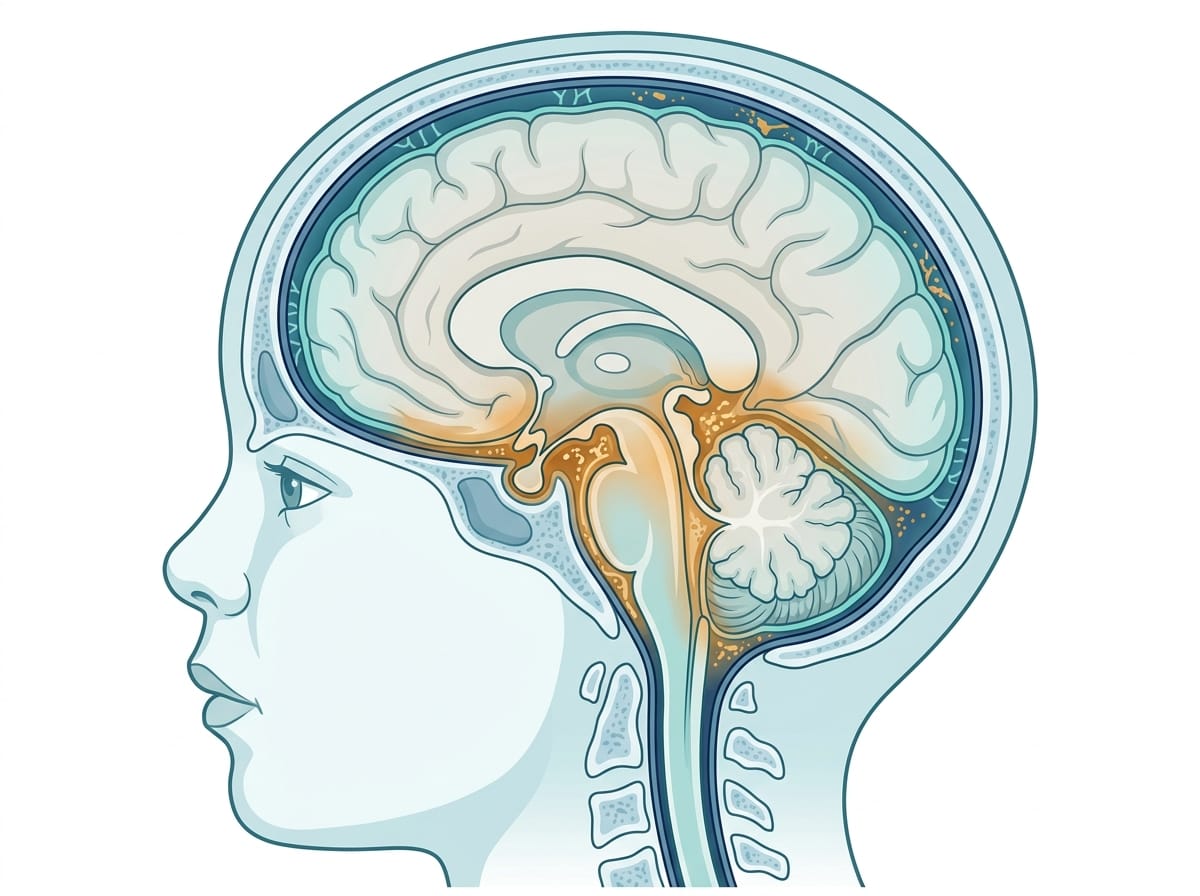
The inflammation at the base of the brain — a thick inflammatory exudate that forms as the immune system responds to TB — obstructs the pathways through which cerebrospinal fluid normally flows and is absorbed. Cerebrospinal fluid (CSF) is produced continuously inside the brain’s ventricles. It flows through specific channels and is absorbed at specific sites around the brain and spinal cord.
When those channels and absorption sites become blocked by TB-related inflammation, CSF cannot drain. It accumulates inside the ventricles. The ventricles expand. Pressure builds inside the skull.
This is communicating hydrocephalus — the most common type associated with TB meningitis. Unlike non-communicating hydrocephalus where a structural blockage prevents CSF flow between chambers, communicating hydrocephalus in TB occurs because the absorption mechanism at the outer surface of the brain is impaired by inflammation.
The distinction matters for treatment. Some surgical approaches work better for one type than the other. Understanding which type of hydrocephalus your child has is a question worth asking your neurosurgical team directly.

The Delayed Diagnosis Problem
TB-related hydrocephalus presents a specific diagnostic challenge that families should understand.
The initial symptoms of TB meningitis in young children — fever, irritability, poor feeding, vomiting — overlap significantly with many other childhood illnesses. Hydrocephalus symptoms that develop as a consequence — head enlargement in infants, lethargy, behavioural changes in toddlers — can appear days or weeks into a course of illness already attributed to another cause.
A child being treated for pulmonary TB who develops neurological symptoms is not always immediately reassessed for brain involvement. The assumption that treatment is working can delay the recognition that a second process — brain inflammation and hydrocephalus — is developing concurrently.
If your child has a confirmed TB diagnosis of any kind and develops any of the following, request urgent neurological assessment:
- Fever that persists or worsens despite TB treatment
- New onset vomiting, particularly in the morning
- Changes in behaviour, alertness, or irritability
- Changes in eye movement or vision
- Seizures
- Headache in a child old enough to report it
These symptoms in the context of known TB should prompt immediate consideration of TB meningitis and its neurological complications including hydrocephalus.
What the Research Shows
A twenty-year review of paediatric tuberculous meningitis by van Well et al., published in Pediatrics, examined outcomes in children with confirmed TB meningitis and found that hydrocephalus was the most common complication — present in a significant proportion of cases. Earlier stages of disease at presentation consistently predicted better neurological outcomes, reinforcing the importance of early recognition. Available at PubMed.
A comprehensive review of central nervous system tuberculosis by Rock et al., published in Clinical Microbiology Reviews, described the pathophysiology of how TB-related inflammation creates communicating hydrocephalus through obstruction of CSF reabsorption at the basal cisterns — the spaces at the base of the brain where drainage pathways are concentrated. The review notes that this complication can develop rapidly and can be the primary driver of neurological deterioration even when antibiotic treatment of the TB infection is appropriate. Available at PubMed.
Research by Principi and Esposito examining paediatric TB meningitis specifically found that the diagnosis is frequently delayed in children — partly because the early clinical picture is non-specific, and partly because clinicians in settings with lower TB prevalence may not consider it promptly. This delay contributes to the severity of neurological complications at the time of diagnosis. Available at PubMed.
What I want to be honest about: most clinical research on TB meningitis has been conducted in high-prevalence settings — South Asia, Sub-Saharan Africa — and data specific to the youngest children (under two) is more limited than for older age groups. The mechanisms described above are well-established. Their precise application varies with age, immune status, and TB strain.
Treatment — What Families Should Know
Treatment of TB-related hydrocephalus involves addressing both the infection and its neurological consequences.
Anti-TB medication: The standard four-drug regimen (isoniazid, rifampicin, pyrazinamide, ethambutol) targets the TB bacteria. Duration for TB meningitis is typically twelve months — longer than pulmonary TB — because penetration of some drugs into the central nervous system is limited.
Corticosteroids: As discussed in our article on steroids and brain inflammation, corticosteroids — most commonly dexamethasone — are added specifically to reduce the inflammatory response that causes the CSF obstruction leading to hydrocephalus. Clinical trials show they reduce mortality and neurological complications in TB meningitis.
Hydrocephalus management: Depending on severity, hydrocephalus may be managed through:
- Medical management with corticosteroids reducing inflammation and allowing CSF flow to recover
- Serial lumbar punctures to temporarily reduce CSF pressure in certain cases
- Surgical intervention (shunt placement or ETV) if hydrocephalus is severe or does not respond to medical management
Not every child with TB-related hydrocephalus requires surgery. The decision depends on the severity of hydrocephalus, the response to medical treatment, and the clinical picture. This is an important conversation to have explicitly with the neurosurgical and infectious disease teams together.

What This Means for Your Family
If your child has been diagnosed with TB anywhere in the body, ask your treating physician directly: “Is there any risk of TB spreading to the brain, and what signs should prompt us to seek neurological assessment?”
If your child has been diagnosed with both TB and hydrocephalus, ask: “Is the hydrocephalus communicating or non-communicating? Is it related to the TB infection, and how does that affect the treatment approach?”
Understanding the relationship between the two diagnoses — rather than treating them as separate unrelated events — helps families ask better questions, recognise relevant symptoms earlier, and participate more effectively in treatment decisions.
The connection between infection and hydrocephalus in your child is not a medical coincidence. It is a specific, well-understood pathway. Knowing that pathway gives you something to track and something to ask about.
Questions to Ask Your Medical Team
Ask your infectious disease specialist and neurologist together:
- Is my child’s hydrocephalus confirmed to be caused by the TB infection?
- Is this communicating or non-communicating hydrocephalus — and does the type affect the treatment plan?
- Will the hydrocephalus resolve with TB treatment and steroids, or is surgical intervention likely to be needed?
- How long is the anti-TB course for brain TB, and why is it longer than lung TB?
- What neurological follow-up will my child need after completing TB treatment?
- What signs of worsening hydrocephalus should prompt me to return urgently?

Frequently Asked Questions About Brain TB and Hydrocephalus
Can TB cause hydrocephalus in children?
Yes. Tuberculous meningitis — TB infection of the brain’s membranes — causes inflammation that blocks the pathways through which cerebrospinal fluid normally drains. This obstruction leads to fluid accumulation and hydrocephalus. It is one of the most common infectious causes of hydrocephalus in young children globally.
How does TB reach the brain from the lungs?
TB bacteria can enter the bloodstream from the lungs and travel to other body systems — including the brain and its surrounding membranes. This process, called haematogenous dissemination, is more common in young children whose immune systems and blood-brain barrier are less developed. The brain infection can develop before, during, or after lung TB treatment.
Does every child with TB meningitis develop hydrocephalus?
No. Hydrocephalus is a complication of TB meningitis, not an inevitable outcome. The risk is higher with more severe or delayed-diagnosis meningitis. Early treatment with anti-TB medicines and corticosteroids reduces the severity of inflammation and lowers the risk of hydrocephalus developing or progressing.
Can TB-related hydrocephalus resolve without surgery?
In some cases yes. When hydrocephalus results from inflammatory obstruction of CSF drainage — rather than a structural blockage — reducing that inflammation with steroids and treating the underlying infection can allow CSF flow to recover. Whether this is sufficient depends on the severity of hydrocephalus and the individual clinical picture. Surgical intervention is needed when medical management cannot adequately control intracranial pressure.
The first diagnosis was lungs. The second was brain. Nobody sat with us and explained how one had led to the other — how the bacteria had moved, how the inflammation had built, how the fluid had accumulated while we were focused on the treatment that had already started.
Understanding that pathway did not change what had happened. But it changed how we asked questions, how we monitored, how we understood what we were watching for.
If your child has received both diagnoses — or if you are reading this because you are trying to make sense of a child who had TB and then developed something neurological — you are not looking at two separate misfortunes. You are looking at one process.
Knowing what that process is makes it possible to face it with clearer eyes.
This article is for informational purposes only and does not constitute medical advice. Always consult your neurologist, paediatrician, and infectious disease specialist for guidance specific to your child’s situation. Read our full disclaimer: braincarepath.com/disclaimer/
Bibliography
- van Well GT, Paes BF, Terwee CB, et al. Twenty years of pediatric tuberculous meningitis: a retrospective cohort study in the western cape of South Africa. Pediatrics. 2009;123(1):e1-8. Available at PubMed.
- Rock RB, Olin M, Baker CA, Molitor TW, Peterson PK. Central nervous system tuberculosis: pathogenesis and clinical aspects. Clin Microbiol Rev. 2008;21(2):243-261. Available at PubMed.
- Principi N, Esposito S. Diagnosis and therapy of tuberculous meningitis in children. Tuberculosis. 2012;92(5):377-383. Available at PubMed.
- Thwaites GE, van Toorn R, Schoeman J. Tuberculous meningitis: more questions, still too few answers. Lancet Neurol. 2013;12(10):999-1010. Available at PubMed.
Frequently Asked Questions About TB and the Brain
Can tuberculosis affect the brain?
Yes. Tuberculosis can infect the membranes covering the brain, causing tuberculous meningitis, or form mass lesions called tuberculomas. CNS tuberculosis is among the most serious forms of TB and is more common in regions where TB remains widespread, including South Asia.
How does TB cause hydrocephalus?
Tuberculous meningitis produces thick inflammatory exudate at the base of the brain. This blocks the normal flow and absorption of cerebrospinal fluid, so fluid accumulates and pressure rises. Hydrocephalus is one of the commonest complications of TB meningitis.
What are the symptoms of TB in the brain?
Early symptoms are often vague — persistent low-grade fever, weight loss, night sweats, headache and irritability over weeks rather than days. Later signs include vomiting, neck stiffness, confusion, seizures, and reduced consciousness. The slow onset frequently delays diagnosis.
Is brain TB curable?
It is treatable, and many people recover, but outcome depends heavily on how early treatment starts. Treatment involves prolonged anti-tuberculous therapy, usually 9 to 12 months, sometimes with steroids. Hydrocephalus may additionally require a shunt.