

Post-Infectious Hydrocephalus: Vital Causes and Full Recovery
By Haris Bin Tahir
Father of a hydrocephalus survivor. Independent researcher. Not a doctor.
Founder, Brain Care Path · braincarepath.com
Quick answer: Post-infectious hydrocephalus — hydrocephalus caused by bacterial meningitis, TB meningitis, or viral brain infection — accounts for a significant proportion of acquired hydrocephalus in children and is distinct in its mechanism, treatment, and recovery trajectory from congenital forms.
Key Takeaways
- Post-infectious hydrocephalus — hydrocephalus caused by bacterial meningitis, TB meningitis, or viral brain infection — accounts for a significant proportion of acquired hydrocephalus in children and is distinct in its mechanism, treatment, and recovery trajectory from congenital forms
- The inflammation left behind by brain infection physically blocks the pathways through which cerebrospinal fluid normally drains — meaning treatment must address both the infection and its neurological consequences simultaneously
- Recovery in post-infectious hydrocephalus is frequently better than in other acquired forms because the underlying brain architecture, prior to the infection, was intact — giving neuroplasticity a stronger foundation to work from
The infection was the first crisis. But post-infectious hydrocephalus — the condition that follows — is often the longer one. Everyone focused on the fever chart, the cultures, the antibiotic levels.
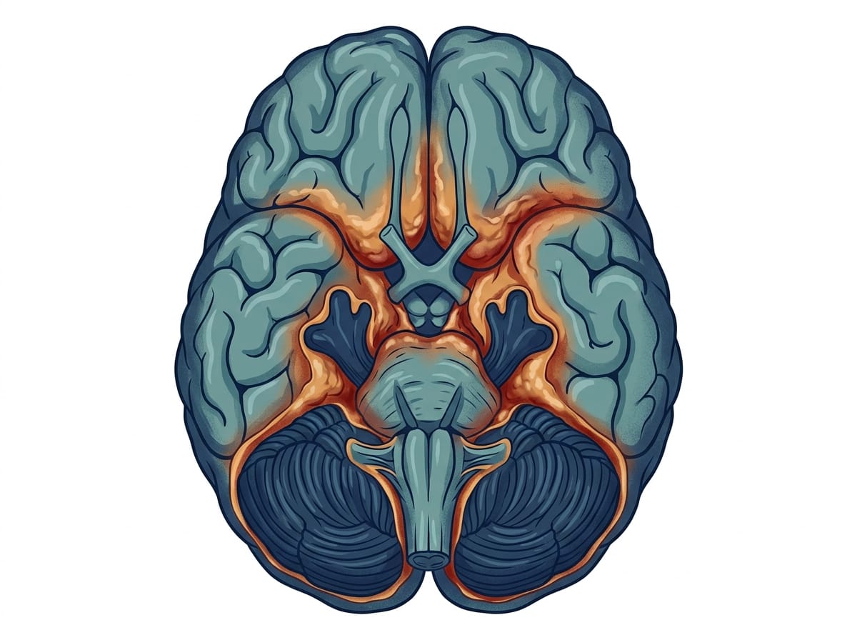
But infection in the brain does not leave cleanly. It leaves something behind: a residue of inflammation and scar tissue at the base of the skull where the meninges had been fighting. And it was this residue that changed the drainage architecture of his brain — that blocked the channels through which cerebrospinal fluid had been flowing freely since before he was born.
The infection became controlled. The hydrocephalus it created did not resolve by itself.
Post-infectious hydrocephalus is one of the most common acquired forms of hydrocephalus in children worldwide — and one of the least explained to families who receive it as a secondary diagnosis after an already devastating primary one. This article addresses what it is, how it differs from other forms, and what full recovery genuinely looks like.
What Post-Infectious Hydrocephalus Is
Post-infectious hydrocephalus develops as a consequence of an infection that has reached and inflamed the meninges — the membranes surrounding the brain and spinal cord — or the brain tissue itself.
The infections most commonly responsible in children include:
Bacterial meningitis — caused by bacteria such as Streptococcus pneumoniae, Neisseria meningitidis, or Haemophilus influenzae. Bacterial meningitis carries the highest risk of post-infectious hydrocephalus among common childhood infections.
Tuberculous meningitis (TB meningitis) — caused by Mycobacterium tuberculosis reaching the meninges. TB meningitis produces a particularly thick inflammatory exudate at the base of the brain that is strongly associated with CSF drainage obstruction.
Viral meningoencephalitis — caused by various viruses. Generally lower risk of hydrocephalus than bacterial or TB forms, but can cause it in severe cases.
Neonatal sepsis and intraventricular haemorrhage — in premature infants, bleeding within the ventricles can produce a post-haemorrhagic hydrocephalus that shares mechanism and clinical features with post-infectious forms.
How It Differs From Congenital Hydrocephalus
Congenital hydrocephalus is present from before birth — caused by developmental abnormalities, genetic factors, or structural issues in the brain’s drainage pathways.
Post-infectious hydrocephalus develops in a brain that was previously normal. The drainage pathways were functioning. The brain architecture was intact. The infection disrupted a working system rather than developing in the context of one that never worked correctly.
This distinction has significant implications for prognosis. A child whose brain was developing normally before the infection has a different neurological foundation for recovery than one with a congenital structural abnormality. The damage in post-infectious hydrocephalus is real — but it is imposed on a previously normal architecture, and the brain’s capacity to recover is often correspondingly stronger.
Key Takeaways
- Congenital hydrocephalus is present from birth due to developmental or genetic factors.
- In mild cases where the infection is rapidly controlled and inflammation is minimal, some degree of spontaneous CSF drainage recovery can occur.
- There is no single timeline.
- Yes.
The Mechanism — How Infection Creates Hydrocephalus
The pathway from brain infection to hydrocephalus follows a consistent biological sequence.
When bacteria or TB bacilli reach the meninges, the immune system responds with inflammation — releasing immune cells and inflammatory proteins into the subarachnoid space (the fluid-filled space between two of the meningeal layers). This inflammatory response is necessary to fight the infection. But it also produces physical changes to the meningeal tissue.
The most critical site of damage is the basal cisterns — a group of CSF spaces at the base of the brain where drainage pathways converge. In TB meningitis specifically, a dense inflammatory mass called a basal exudate forms here. This exudate has a thick, fibrinous consistency that physically obstructs CSF flow.
As the infection is treated and inflammation partially resolves, this exudate can leave behind fibrous scar tissue. The scarring narrows or blocks the channels and absorption sites that CSF requires. Even though the active infection is gone, the obstruction it created persists.
This is why post-infectious hydrocephalus can develop or worsen even as the infection itself improves — and why families sometimes find themselves confused when their child appears to be responding to treatment but neurological symptoms are worsening.

Diagnosis — How It Is Confirmed
Post-infectious hydrocephalus is typically identified through brain imaging — MRI or CT scan — showing enlarged ventricles in the context of a known or suspected brain infection.
MRI is preferred because it provides detailed information about the degree of ventricular enlargement, the presence and extent of basal exudate or scarring, the involvement of brain parenchyma (brain tissue itself), and the pattern of hydrocephalus — communicating versus non-communicating.
Lumbar puncture (spinal tap) may be performed to measure CSF pressure, examine CSF composition, and culture for the causative organism. Serial lumbar punctures are sometimes used as a temporary measure to reduce CSF pressure while the underlying infection is being controlled.
CSF opening pressure on lumbar puncture provides direct measurement of intracranial pressure — a more accurate indicator of the severity of hydrocephalus than imaging alone.

Treatment — Addressing Both the Infection and the Hydrocephalus
Treatment of post-infectious hydrocephalus operates on two simultaneous tracks.
Track 1 — Treating the Underlying Infection
The causative infection must be treated aggressively and completely. For bacterial meningitis, this means intravenous antibiotics — typically for 10-21 days depending on the organism and clinical response. For TB meningitis, this means the full anti-TB drug regimen — typically 12 months for CNS involvement.
Corticosteroids are added specifically to reduce meningeal inflammation. Clinical trials have demonstrated that dexamethasone significantly reduces mortality and neurological complications in both bacterial and tuberculous meningitis when started early and maintained appropriately.
Track 2 — Managing the Hydrocephalus
Medical management: In some cases — particularly when the underlying infection is well-controlled and hydrocephalus is moderate — the hydrocephalus can be monitored carefully while infection treatment proceeds. As inflammation reduces with treatment, some degree of CSF drainage recovery can occur. This approach requires frequent clinical monitoring and readiness to escalate to surgical management if needed.
Serial lumbar punctures: Repeated lumbar punctures can temporarily reduce CSF pressure and provide clinical relief during the acute period. This approach is used selectively — it is not a permanent solution, but it can buy time for the infection to respond to treatment.
Surgical management: When hydrocephalus is severe, progressing, or not responding to medical management, surgical intervention is considered. Options include VP shunt placement, external ventricular drainage (a temporary measure in acute settings), or endoscopic third ventriculostomy (ETV) where appropriate.
Not every child with post-infectious hydrocephalus requires surgery. The decision depends on the type and severity of hydrocephalus, the response to treatment, and the clinical trajectory — and it is best made by a team including neurosurgery and the infectious disease or paediatric neurology team together.

What the Research Shows
A twenty-year retrospective study of paediatric tuberculous meningitis by van Well et al., published in Pediatrics, found that hydrocephalus was the most common neurological complication — present in over half of cases with severe disease. Outcomes were significantly better in children who received early diagnosis and combined anti-TB and steroid treatment.
Research on post-infectious hydrocephalus in sub-Saharan Africa by Warf et al., published in the Journal of Neurosurgery, examined outcomes specifically in infants with post-infectious hydrocephalus versus congenital forms. The study found that post-infectious hydrocephalus had better cognitive outcomes than some congenital forms — consistent with the hypothesis that a previously normal brain architecture provides a stronger platform for recovery.
Research consistently identifies the timing of hydrocephalus management as a key determinant of outcome. Prolonged elevated intracranial pressure in the post-infectious period — even after the infection itself is controlled — produces ongoing white matter injury. Monitoring and treating the hydrocephalus component is as important as treating the infection.
Recovery — What Full Recovery Means
Recovery in post-infectious hydrocephalus is real. It is also non-linear and individual.
What full recovery does not mean is an absence of any lasting effect. The infection and the pressure it caused have affected the brain during a sensitive developmental period. Some children achieve outcomes that are indistinguishable from their peers within two to three years. Others carry lasting difficulties in specific domains — learning, attention, motor precision — that require targeted support.
What full recovery means, more accurately, is the full expression of the brain’s adaptive capacity given the nature and extent of injury. That capacity is significantly greater than most families are led to believe at the point of diagnosis.
The strongest predictors of better recovery in post-infectious hydrocephalus are younger age at infection (higher neuroplasticity available), shorter duration of elevated pressure before effective treatment, consistent and targeted rehabilitation begun early, adequate sleep and nutrition throughout the recovery period, and absence of recurrent infection or untreated seizures.

Questions to Ask Your Child’s Medical Team
- Is my child’s hydrocephalus confirmed to be post-infectious — and which infection caused it?
- Is this communicating or non-communicating hydrocephalus, and does the type affect the treatment plan?
- What is the current intracranial pressure measurement, and is it trending upward, stable, or downward?
- At what point will you consider surgical management, and what would that decision be based on?
- What monitoring plan is in place for the coming weeks and months?
- Given the type and severity, what is a realistic expectation for neurological recovery?
Frequently Asked Questions
What is the difference between post-infectious and congenital hydrocephalus?
Congenital hydrocephalus is present from birth due to developmental or genetic factors. Post-infectious hydrocephalus develops after birth as a result of brain infection — it affects a brain that was previously developing normally. This distinction affects prognosis: post-infectious hydrocephalus often has stronger recovery potential because the underlying brain architecture was intact before the infection.
Can post-infectious hydrocephalus resolve on its own?
In mild cases where the infection is rapidly controlled and inflammation is minimal, some degree of spontaneous CSF drainage recovery can occur. In most moderate to severe cases, the fibrous scarring left by the infection’s inflammatory response creates persistent CSF drainage obstruction that requires active management — medical, surgical, or both.
How long does post-infectious hydrocephalus take to resolve?
There is no single timeline. The active infection must first be fully treated — which for TB meningitis means a twelve-month course. The hydrocephalus itself may require ongoing management throughout and after this period. Neurological recovery from the combined effects of infection and pressure continues over months to years.
Is post-infectious hydrocephalus more common in certain infections?
Yes. Bacterial meningitis and TB meningitis carry the highest risk of hydrocephalus as a complication. TB meningitis specifically is associated with the formation of a basal exudate that is strongly hydrocephalus-producing. Viral meningitis generally carries lower risk. Intraventricular haemorrhage in premature infants produces a closely related condition called post-haemorrhagic hydrocephalus.
The infection became controlled. The hydrocephalus it had created took longer.
Those were different problems requiring different treatments, different monitoring, and different types of vigilance. Understanding that they were connected but distinct was one of the most useful pieces of knowledge we found — because it stopped us from expecting the hydrocephalus to resolve when the infection resolved, and it helped us ask the right questions at the right appointments.
The infection was the cause. The hydrocephalus was the consequence. Recovery from the consequence required its own time, its own treatment, and its own patience.
This article is for informational purposes only and does not constitute medical advice. Always consult your neurologist, paediatrician, and infectious disease specialist for guidance specific to your child’s situation. Read our full disclaimer: braincarepath.com/disclaimer/
Medically Reviewed by Dr. Abubakar Siddique, MBBS, FCPS (Neurology) — Consultant Interventional Neurologist, HOD Neurology and Stroke Unit, Akhter Saeed Medical and Dental College, Lahore.
Bibliography
- van Well GT, Paes BF, Terwee CB, et al. Twenty years of pediatric tuberculous meningitis. Pediatrics. 2009;123(1):e1-8. Available at: https://pubmed.ncbi.nlm.nih.gov/19171623/
- Warf BC. Hydrocephalus in Uganda: the predominance of infectious origin and primary management with endoscopic third ventriculostomy. J Neurosurg. 2005;102(1 Suppl):1-15. Available at: https://pubmed.ncbi.nlm.nih.gov/15926703/
- Thwaites GE, Nguyen DB, Nguyen HD, et al. Dexamethasone for the treatment of tuberculous meningitis in adolescents and adults. N Engl J Med. 2004;351(17):1741-1751. Available at: https://pubmed.ncbi.nlm.nih.gov/15577987/
- Isaacs AM, Riva-Cambrin J, Yavin D, et al. Age-specific global epidemiology of hydrocephalus. PLoS One. 2018;13(10):e0204926. Available at: https://pubmed.ncbi.nlm.nih.gov/30286167/