

Communicating Hydrocephalus: What the Diagnosis Truly Means
Quick answer: Communicating hydrocephalus means cerebrospinal fluid (CSF) still flows freely between all the brain’s ventricles, but the body cannot absorb it fast enough. Fluid builds up and raises pressure. It differs from non-communicating hydrocephalus, where a physical blockage stops CSF moving between chambers.
Key Takeaways
- Hydrocephalus is divided into two broad types — communicating and non-communicating — and the distinction directly affects which treatment approach is most appropriate
- Communicating hydrocephalus means cerebrospinal fluid can still move between the brain’s chambers but cannot be adequately absorbed at the outer surface — it is the type most commonly caused by infection, bleeding, or inflammation
- Understanding which type your child has is not a minor detail — it shapes whether ETV surgery is an option, which shunt settings work best, and what the long-term monitoring should look like
The doctor used the word communicating as though it explained something.
He wrote it on a piece of paper — communicating hydrocephalus — and slid it across the desk. I looked at it. I had expected the word to clarify something. Instead I had a new term I did not understand sitting next to a diagnosis I was only beginning to process.
What was communicating? Communicating with what? Why did the type matter? Was it better or worse than the other kind?
Nobody took the time to answer those questions in that consultation room. The assumption, I think, was that the diagnosis itself was enough to manage. The classification was for the clinicians.
It is not only for the clinicians. Understanding what type of hydrocephalus your child has changes how you ask questions, how you evaluate treatment options, and how you understand the monitoring that follows. This article explains what the distinction actually means.
The Two Types — What They Mean in Plain Language
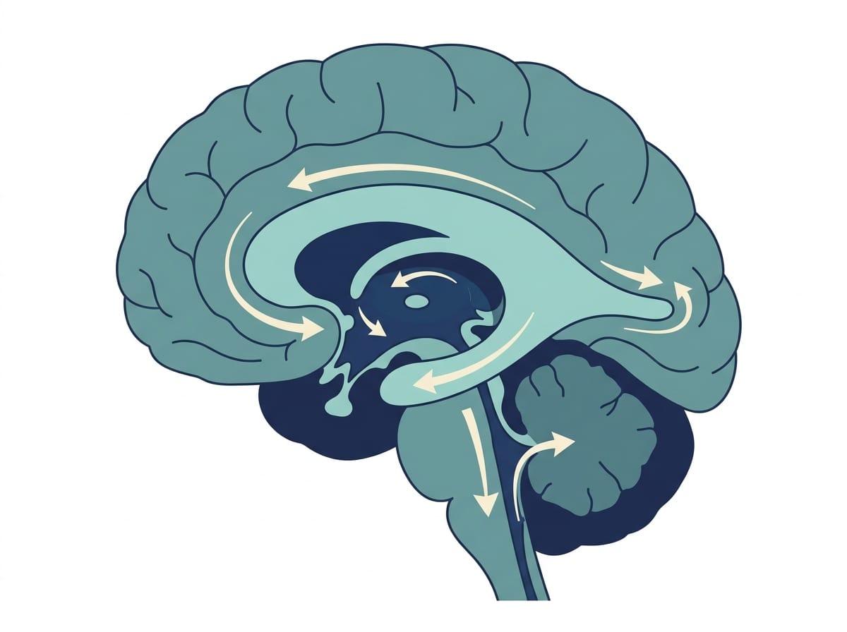
The brain produces cerebrospinal fluid — a clear liquid that circulates through chambers inside the brain called ventricles, flows through specific connecting passages, moves into the subarachnoid space surrounding the brain and spinal cord, and is finally absorbed into the bloodstream at specialised sites called arachnoid granulations.
This is a continuous cycle. CSF is produced, flows, and is absorbed — constantly, day and night.
Hydrocephalus occurs when this cycle breaks down. But it can break down in two fundamentally different places.
Non-Communicating Hydrocephalus
In non-communicating hydrocephalus — also called obstructive hydrocephalus — there is a physical blockage somewhere within the ventricular system itself. CSF cannot flow between chambers because something is blocking the passage.
The most common site is the aqueduct of Sylvius — a narrow channel connecting the third and fourth ventricles. When this narrows or is completely blocked, the ventricles above the blockage expand. The ventricles below it remain normal size.
The blockage can be caused by a tumour, a cyst, a congenital narrowing called aqueductal stenosis, scarring from a previous bleed, or compression from surrounding tissue.
Communicating Hydrocephalus
In communicating hydrocephalus, CSF can still move freely between all the ventricles — all chambers are communicating with each other, hence the name. The problem is not inside the ventricles. It is at the absorption site.
CSF reaches the outer surface of the brain but cannot be adequately absorbed into the bloodstream. The arachnoid granulations — the absorption structures — are impaired. Fluid accumulates because the exit route is blocked or insufficient, not because the internal pathways are obstructed.
All four ventricles typically enlarge together, because the pressure backs up throughout the entire ventricular system rather than above a specific point.
What Causes Communicating Hydrocephalus
Understanding the cause of communicating hydrocephalus helps families understand both the treatment and the prognosis.
Infection and Inflammation
This is the most relevant cause for our family and for many families reading this site.
When bacterial meningitis, TB meningitis, or viral meningoencephalitis inflames the meninges — the membranes surrounding the brain — it can damage or block the arachnoid granulations responsible for CSF absorption. Even after the infection is effectively treated, this damage can persist as fibrous scarring that permanently impairs drainage.
This is post-infectious communicating hydrocephalus. The ventricles were communicating normally before the infection. The infection damaged the absorption mechanism. The result is persistent hydrocephalus even when the infection itself is gone.
Intraventricular Haemorrhage
Bleeding within the ventricles — most commonly in premature infants — can deposit blood products at the arachnoid granulations, impairing their function. Post-haemorrhagic hydrocephalus is a form of communicating hydrocephalus with this mechanism.
Subarachnoid Haemorrhage
Bleeding in the subarachnoid space — from trauma, a ruptured aneurysm, or other causes — can similarly impair CSF absorption at the granulations, producing communicating hydrocephalus as a complication.
Congenital and Idiopathic
Some communicating hydrocephalus is present from birth without an identifiable infectious or haemorrhagic cause. Developmental differences in the arachnoid granulations or in CSF production can produce communicating hydrocephalus without a precipitating event.

Why the Distinction Matters for Treatment
The type of hydrocephalus significantly affects which treatments are most appropriate.
ETV — Endoscopic Third Ventriculostomy
ETV creates a new CSF drainage pathway by making a small opening in the floor of the third ventricle, bypassing the obstructed aqueduct and allowing CSF to flow directly into the subarachnoid space.
ETV works well for non-communicating hydrocephalus — specifically aqueductal stenosis — because it bypasses the internal blockage.
ETV is generally less effective for communicating hydrocephalus, because the problem is not an internal blockage but impaired absorption at the outer surface. Creating a new pathway into the subarachnoid space does not help if absorption there is already impaired.
This is one of the most clinically important implications of the communicating/non-communicating distinction. Families whose child has communicating hydrocephalus should ask their neurosurgeon directly whether ETV is appropriate for their specific type — and what the evidence shows for ETV success rates in post-infectious communicating hydrocephalus specifically.
VP Shunt
A VP shunt bypasses the entire natural CSF pathway — draining fluid from the ventricles directly to the peritoneal cavity, where it is absorbed independent of the impaired arachnoid granulations.
VP shunts work for both types of hydrocephalus — they bypass the problem regardless of where the problem is located. For communicating hydrocephalus, shunting is often the primary surgical option because ETV is less reliable.
Medical Management
In specific cases of communicating hydrocephalus — particularly post-infectious types where the underlying inflammation is being actively treated — medical management with corticosteroids can allow some recovery of arachnoid granulation function. This is not appropriate for all cases but is a genuine option in specific clinical contexts, particularly when infection is the causative factor and is being treated aggressively.

Communicating vs Non-Communicating Hydrocephalus: A Comparison
| Feature | Communicating | Non-Communicating |
|---|---|---|
| CSF flow | Flows freely between ventricles | Blocked inside the ventricular system |
| Core problem | Absorption failure | Physical obstruction |
| Ventricles affected | Usually all, fairly evenly | Only those above the blockage |
| Common causes | Meningitis, haemorrhage, head injury | Aqueductal stenosis, tumour, cyst |
| Usual treatment | VP shunt | ETV often possible |
| Is ETV suitable? | Usually not — bypassing a blockage does not fix absorption | Often yes |
How Communicating Hydrocephalus Develops: The Pathophysiology
Your brain produces roughly 500 ml of cerebrospinal fluid every day. Because the total volume held at any moment is only about 150 ml, the entire supply is replaced several times daily. Production and absorption must therefore stay closely balanced.
Absorption happens mainly through the arachnoid granulations — small structures on the brain’s surface that pass CSF back into the bloodstream. In communicating hydrocephalus, these granulations become scarred, inflamed, or clogged. Consequently, fluid keeps arriving but cannot leave quickly enough.
This explains a detail that confuses many families: the plumbing inside the brain is open. Nothing is physically blocking the ventricles. The problem sits at the drain, not the pipes.
How Is Communicating Hydrocephalus Treated?
Treatment usually means diverting fluid, because there is currently no reliable way to repair damaged absorption sites.
- Ventriculoperitoneal (VP) shunt — the most common approach. A thin tube carries CSF from a ventricle to the abdomen, where the body absorbs it. Read our VP shunt surgery guide.
- Ventriculoatrial or ventriculopleural shunt — used when the abdomen is unsuitable, for example after repeated infection or scarring.
- Endoscopic third ventriculostomy (ETV) — generally less effective here. ETV creates a bypass around a blockage, and in communicating hydrocephalus there is no blockage to bypass. See our ETV outcomes research summary.
- Treating the underlying cause — for instance clearing an infection. This occasionally reduces or resolves the hydrocephalus, particularly in post-infectious cases.
Some adults with normal pressure hydrocephalus undergo a lumbar drain trial first. If walking or thinking improves temporarily, a shunt is more likely to help. Additionally, this trial gives families useful evidence before committing to surgery.
What the Research Shows
A contemporary classification framework for hydrocephalus published by Rekate in Seminars in Pediatric Neurology proposed moving beyond the simple communicating/non-communicating binary toward a more mechanistic understanding of CSF dynamics. The paper emphasised that treatment decisions should be based on the underlying mechanism — not just the structural classification — and that the same clinical presentation can have fundamentally different causes requiring different approaches. Available at: https://pubmed.ncbi.nlm.nih.gov/19660706/
Research by Kahle et al. in the Lancet confirmed that the cause and mechanism of hydrocephalus are the primary determinants of treatment selection and long-term outcome — more so than the degree of ventricular enlargement on imaging alone. Post-infectious communicating hydrocephalus was identified as having outcomes that varied substantially based on the severity of the underlying infection and the timing of management. Available at: https://pubmed.ncbi.nlm.nih.gov/26256071/
I want to be honest about something the research does not fully resolve: the long-term outcomes for communicating hydrocephalus caused by infection are more variable than those for surgically corrected aqueductal stenosis. The infectious damage to absorption mechanisms can be partial or complete, and its reversibility depends on factors that are not always predictable at the time of diagnosis.
What This Means for Your Family
Ask your neurosurgeon directly and clearly: is my child’s hydrocephalus communicating or non-communicating? What is the probable cause? Does the type affect whether ETV or a shunt is the better option for my child specifically?
If the answer is communicating hydrocephalus caused by infection — the follow-up questions are:
- Is there any remaining active inflammation that aggressive treatment could reduce?
- Is the absorption impairment likely to be permanent or potentially reversible with treatment?
- If a shunt is placed, what type and setting is most appropriate for communicating hydrocephalus?
The type is not a minor classification detail. It is a clinically meaningful distinction that should be part of every treatment conversation.

Questions to Ask Your Neurosurgeon
- Is my child’s hydrocephalus communicating or non-communicating — and how was this determined?
- What is the most likely underlying cause of the CSF drainage problem in my child’s case?
- Does the type affect whether ETV or VP shunting is more appropriate?
- If communicating, is there any evidence that the absorption impairment could partially recover with treatment of the underlying cause?
- What monitoring is appropriate given the specific type and cause of hydrocephalus?
- Will you repeat imaging to assess whether the type or degree of hydrocephalus has changed with treatment?
Frequently Asked Questions
What is the difference between communicating and non-communicating hydrocephalus?
In communicating hydrocephalus, CSF flows freely between all brain chambers but cannot be adequately absorbed at the outer surface of the brain. In non-communicating hydrocephalus, a physical blockage within the ventricular system prevents CSF from flowing between chambers. The distinction affects which surgical treatments are most appropriate and what the likely cause is.
Is communicating hydrocephalus more serious than non-communicating?
Neither type is inherently more serious than the other. Outcomes depend more on the underlying cause, the timing of diagnosis, and the effectiveness of treatment than on the type classification. Communicating hydrocephalus caused by infection or haemorrhage may have more complex outcomes because of the concurrent brain injury from those events.
Can communicating hydrocephalus resolve on its own?
In mild cases where the underlying cause — such as meningeal inflammation — responds well to treatment and the absorption impairment is partial rather than complete, some degree of spontaneous improvement can occur. Complete resolution without intervention is uncommon in established communicating hydrocephalus. Medical management with steroids and treatment of the underlying infection can support recovery in specific cases.
Why does communicating hydrocephalus affect all ventricles equally?
Because the problem is at the absorption site rather than at an internal blockage point. When all ventricles can communicate freely but fluid cannot exit the system at the outer surface, pressure backs up equally throughout the entire ventricular system — causing all chambers to enlarge together, rather than only those above a specific blockage point.
The word communicating appeared on a piece of paper and I did not know what it meant.
What it meant, I learned over the following months, was that the pathways inside my son’s brain were intact. The damage was at the outer surface — the absorption sites left scarred by the infection. The treatment had to account for that specific mechanism.
Understanding the type changed how we asked questions at every subsequent appointment. It changed what we listened for in the answers. It changed what we pushed back on when the recommendations did not seem to account for the specific nature of what his brain was dealing with.
The type matters. Ask about it. Keep asking until you understand it.
This article is for informational purposes only and does not constitute medical advice. Always consult your child’s neurosurgeon and neurologist for guidance specific to their diagnosis. Read our full disclaimer: braincarepath.com/disclaimer/
What causes communicating hydrocephalus?
The most common causes are meningitis, bleeding around the brain (subarachnoid or intraventricular haemorrhage), head injury, and previous brain surgery. Each can scar or inflame the arachnoid granulations that absorb cerebrospinal fluid. In some cases, particularly in older adults, no clear cause is ever identified.
How is communicating hydrocephalus treated?
Most people are treated with a ventriculoperitoneal (VP) shunt, which drains excess fluid from the brain to the abdomen. Endoscopic third ventriculostomy is usually less suitable, because it bypasses blockages rather than improving absorption. Treating an underlying infection can sometimes help.
Is communicating hydrocephalus curable?
It is usually managed rather than cured. A shunt controls pressure effectively and many people live full, ordinary lives with one. Occasionally, when hydrocephalus follows an infection that fully resolves, absorption recovers and no long-term treatment is needed.
What is the life expectancy with communicating hydrocephalus?
With effective treatment, most people have a normal or near-normal life expectancy. Outcomes depend far more on the underlying cause and on avoiding complications such as repeated shunt revisions than on the hydrocephalus itself. Read more in our life expectancy research summary.
Bibliography
- Rekate HL. A contemporary definition and classification of hydrocephalus. Semin Pediatr Neurol. 2009;16(1):9-15. Available at: https://pubmed.ncbi.nlm.nih.gov/19260980/
- Kahle KT, Kulkarni AV, Limbrick DD Jr, Warf BC. Hydrocephalus in children. Lancet. 2016;387(10020):788-799. Available at: https://pubmed.ncbi.nlm.nih.gov/26256071/
- Oi S, Di Rocco C. Proposal of “evolution theory in cerebrospinal fluid dynamics.” Childs Nerv Syst. 2006;22(6):589-595. Available at: https://pubmed.ncbi.nlm.nih.gov/16570167/
- Isaacs AM, Riva-Cambrin J, Yavin D, et al. Age-specific global epidemiology of hydrocephalus. PLoS One. 2018;13(10):e0204926. Available at: https://pubmed.ncbi.nlm.nih.gov/30286167/