

Hydrocephalus Diagnosis: How CT, MRI and Ultrasound Truly Work
Quick answer: Hydrocephalus is diagnosed by imaging that shows enlarged ventricles. A CT scan is fastest and used in emergencies. MRI gives the most detail and shows why fluid is building up. Cranial ultrasound is used in babies whose soft spot is still open.
Key Takeaways
- Three main imaging tools are used to diagnose and monitor hydrocephalus — cranial ultrasound, CT scan, and MRI — each with specific strengths, limitations, and appropriate uses depending on the child’s age and clinical situation
- The ventricular measurements shown on imaging reports are more meaningful when understood in context — a parent who knows what the numbers mean can ask better questions and track changes more accurately over time
- MRI is the gold standard for detailed brain assessment in hydrocephalus but is not always the first-line investigation — understanding why one test was chosen over another is a question worth asking at every imaging appointment
They took him for the scan at half past eleven at night.
I sat in a plastic chair outside the imaging department and listened to the sound of the machine through the wall — a repetitive mechanical rhythm that told me something was happening but nothing about what it was finding.
Forty minutes later a radiologist handed a report to the nurse. Nobody explained it to me until the following morning. By then I had read every word of the report three times, understanding perhaps a quarter of it. The words ventricular dilation and transependymal CSF flow appeared several times. I did not know what they meant. I did not know whether the numbers were alarming or expected.
Hydrocephalus diagnosis depends on imaging. Every family navigating this condition encounters scans, reports, and measurements. This article explains what each type of imaging involves, what it is showing, and how to make sense of what the reports say.
The Three Main Imaging Tools
Cranial Ultrasound — First in Young Infants
Cranial ultrasound uses sound waves to create images of the brain through the anterior fontanelle — the soft spot on the top of an infant’s head that remains open until around eighteen months of age.
Because it requires no sedation, no radiation, and can be performed at the bedside, cranial ultrasound is the first-line investigation for suspected hydrocephalus in young infants. It can be repeated frequently as a monitoring tool without the risks associated with radiation exposure from CT scanning.
What ultrasound shows well:
- Ventricular size and shape
- Obvious haemorrhage within the ventricles
- Gross structural abnormalities
- Changes in ventricular size over time when used serially
What ultrasound shows less well:
- Fine detail of brain tissue
- The posterior fossa (back of the brain)
- The cause of hydrocephalus in most cases
- Subtle changes in white matter
Once the fontanelle closes — typically around eighteen months — cranial ultrasound is no longer useful as a brain imaging tool.
CT Scan — Fast, Widely Available, High Radiation
Computed tomography uses X-rays from multiple angles to create cross-sectional images of the brain. In the emergency setting, CT scanning is often the first investigation for a child presenting acutely — because it is fast, widely available around the clock, and highly effective at identifying the most urgent findings.
What CT shows well:
- Ventricular size — CT is excellent at showing ventricular enlargement
- Acute haemorrhage — blood appears bright white on CT immediately after a bleed
- Gross structural abnormalities
- Shunt hardware and its position
- Signs of acute raised intracranial pressure
What CT shows less well:
- Subtle brain tissue changes
- White matter injury
- Posterior fossa detail (bone artefact affects this region)
- The underlying cause of hydrocephalus in many cases
The radiation concern:
CT scans use ionising radiation. In children — whose developing tissues are more sensitive to radiation than adults — this is a genuine consideration. For an acute clinical question where the result will change management immediately, CT is appropriate and justified. For routine monitoring in a stable child, MRI is preferred because it involves no radiation.
Families should feel comfortable asking: is a CT scan necessary at this appointment, or would an MRI be more appropriate given that this is monitoring rather than an emergency assessment?

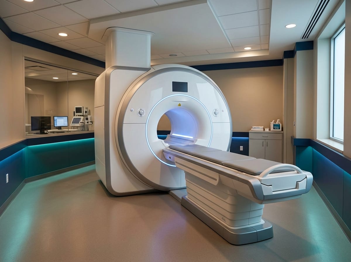
MRI — Most Detailed, No Radiation, Requires Stillness
Magnetic resonance imaging uses magnetic fields and radio waves rather than radiation to produce highly detailed images of brain structure. It is the gold standard investigation for hydrocephalus assessment and carries no radiation risk.
What MRI shows well:
- Detailed brain tissue including white matter changes
- The cause of hydrocephalus in many cases — identifying structural abnormalities, tumours, aqueductal stenosis
- Periventricular white matter injury from pressure
- CSF flow — specialised MRI sequences can visualise CSF movement through the ventricular system
- Fine posterior fossa detail
- All aspects of shunt assessment when clinically indicated
What MRI shows less well:
- Shunt hardware — metal components cause artefact (always inform radiology staff about shunt type and whether it is programmable before any MRI)
- Acute haemorrhage in the very early phase
The sedation challenge:
MRI requires the patient to lie completely still for 20–45 minutes inside a narrow tube while loud mechanical sounds occur. Young children and infants almost always require sedation or general anaesthesia for diagnostic MRI. This adds procedural risk and complexity. In older children who can cooperate, MRI without sedation is often possible.
CT vs MRI vs Ultrasound for Hydrocephalus: Which Scan and When
| CT Scan | MRI | Ultrasound | |
|---|---|---|---|
| Time taken | Under 5 minutes | 30–60 minutes | 5–10 minutes |
| Radiation | Yes | None | None |
| Sedation needed? | Rarely | Often in young children | No |
| Shows the cause? | Sometimes | Usually — best detail | Limited |
| Typical use | Emergency, suspected shunt failure | Planned diagnosis, surgical planning | Babies with an open fontanelle |
| Age suited to | Any age | Any age | Usually under 12 months |
Many hospitals now use a rapid-sequence MRI for children who need repeat imaging. Because it avoids radiation and often avoids sedation, it has become the preferred way to monitor a child over years rather than months.
Understanding the Numbers on Imaging Reports
Imaging reports on children with hydrocephalus typically include measurements of the ventricular system. These numbers appear alarming in isolation. Understanding what they represent in context is more useful than knowing the absolute figure.
Ventricular Index and Evans’ Ratio
Evans’ ratio is one of the most commonly reported measurements — the ratio of the width of the frontal horns of the lateral ventricles to the maximum width of the inner skull at the same level. A ratio above 0.3 is conventionally used as a threshold for hydrocephalus in adults. In children this threshold is applied differently given developmental variation.
What matters more than the absolute ratio is the trend. A stable Evans’ ratio across serial imaging suggests the hydrocephalus is not progressing. A rising ratio suggests the ventricles are enlarging — which warrants clinical reassessment even if the absolute figure appears only mildly abnormal.
Transependymal CSF Flow
This phrase — which appeared on my son’s report and which I initially found alarming — refers to CSF crossing from the ventricles through the ependymal lining into the surrounding white matter. On imaging it appears as a halo of fluid signal around the ventricles.
Transependymal flow is a sign of elevated intraventricular pressure — the ventricles are under pressure and CSF is being forced through the lining into surrounding tissue. It is a sign of active, pressure-generating hydrocephalus rather than simply enlarged ventricles.
Third and Fourth Ventricle Size
Isolated enlargement of the lateral ventricles with normal third and fourth ventricle size suggests a blockage at or above the aqueduct of Sylvius. Enlargement of all four ventricles suggests communicating hydrocephalus with impaired absorption downstream.

Serial Imaging — Monitoring Over Time
For a child with hydrocephalus, imaging is not a one-time event. Serial imaging — repeated scans at defined intervals — allows the treating team to track whether:
- The ventricular size is stable, improving, or enlarging
- A shunt is continuing to function appropriately
- The underlying cause has changed
- White matter injury is progressing or stable
Frequency of serial imaging varies considerably. A child with stable, treated hydrocephalus might have annual or biannual MRI. A child in an acute phase of treatment may have imaging every few days.
Always ask at each imaging appointment: what are we looking for compared to the last scan? Has anything changed? Is the comparison to the previous images documented in this report?

What the Research Shows
A systematic review and evidence-based guidelines for paediatric hydrocephalus published in the Journal of Neurosurgery: Pediatrics examined imaging protocols across multiple centres and confirmed that MRI is preferred over CT for non-urgent hydrocephalus assessment in children due to superior tissue detail and absence of radiation risk. The review noted that the number of CT scans a child receives over a lifetime of hydrocephalus monitoring is a legitimate clinical concern — and that institutions have moved toward MRI-first protocols where sedation facilities allow. Available at: https://pubmed.ncbi.nlm.nih.gov/25988776/
Research on radiation exposure in paediatric neuroimaging has consistently found that CT-related radiation exposure in childhood carries a small but measurable increased risk of radiation-related harm over a lifetime — proportionally higher in children than adults. The clinical benefit of CT in acute situations is unambiguous. The case for CT in routine monitoring — where MRI is available — is less clear. Available at: https://pubmed.ncbi.nlm.nih.gov/22840000/
What This Means for Your Family
Request a copy of every imaging report. They are your child’s records and you are entitled to them. Keep a file — paper or digital — of every scan, every report, and every change in measurements noted over time.
Before each imaging appointment, ask: which type of scan is being done and why? Is radiation-free imaging possible for this particular assessment?
When the report arrives, ask the radiologist or neurologist to explain any measurements that have changed since the last scan — not just whether things look normal, but what specifically is different and what that difference means.
And if a report uses terms you do not understand — as my son’s first report did — write them down and ask. You are not expected to have a radiology degree. You are entitled to an explanation.

Questions to Ask at Imaging Appointments
- Which type of imaging is being performed and why was this type chosen over alternatives?
- Is this a CT scan — and if so, is CT necessary at this appointment or could MRI be used instead?
- What specific measurements or features are being assessed compared to previous imaging?
- Has anything changed since the last scan — and is any change clinically significant?
- What are the next imaging milestones — when will the next scan be and what will it be looking for?
- If my child has a programmable shunt, does the MRI protocol account for potential valve setting changes?
Frequently Asked Questions
Is CT or MRI better for diagnosing hydrocephalus in children?
MRI provides more detailed information and involves no radiation, making it preferable for non-urgent hydrocephalus assessment. CT is faster, more widely available, and better for acute emergencies — particularly when haemorrhage needs to be rapidly excluded. Many centres use CT in the initial acute assessment and then transition to MRI for ongoing monitoring.
How often does a child with hydrocephalus need brain scans?
Frequency varies with the clinical situation. A child with stable, treated hydrocephalus may have annual MRI monitoring. A child in an acute phase or with a recently placed shunt may have imaging every few days initially, then progressively less frequently as stability is established. Always ask your neurosurgical team what the planned imaging schedule is and what would trigger an unscheduled scan.
Is it safe to have an MRI with a VP shunt?
Most modern VP shunts are MRI-compatible at 1.5 Tesla and 3 Tesla field strengths. However, programmable shunts — those with adjustable valve settings — may have their settings altered by the MRI magnetic field. The valve setting must be checked and if necessary adjusted after every MRI in a child with a programmable shunt. Always inform the radiology team about the shunt type before any MRI.
What does ventricular dilation mean on a brain scan?
Ventricular dilation means the fluid-filled chambers inside the brain are larger than expected for the child’s age. It is a finding consistent with hydrocephalus but not diagnostic on its own — normal variants and other conditions can cause mild ventricular enlargement. The degree of dilation, the associated findings, and the clinical presentation together determine whether treatment is indicated.
I sat in that plastic chair for forty minutes listening to a machine through a wall. The report told me things in language I had not been given the tools to read.
Nobody should navigate that experience without some preparation.
The scans are not mysterious. The measurements are not arbitrary. Each image is telling a specific story about what is happening inside the brain. Learning to read even part of that story — to ask the right questions of the people who can read all of it — changes what you are able to do with the information.
Ask for the reports. Keep them. Compare them. Ask what changed and why.
You deserve to understand what the machine found.
This article is for informational purposes only and does not constitute medical advice. Always consult your child’s radiologist, neurosurgeon, and neurologist for guidance on imaging findings specific to your child. Read our full disclaimer: braincarepath.com/disclaimer/
What does hydrocephalus look like on a CT scan?
On a CT scan, hydrocephalus appears as enlarged dark areas in the centre of the brain — these are the fluid-filled ventricles. Radiologists also look for rounded ventricle edges, flattened brain folds pressed against the skull, and a bright rim around the ventricles suggesting fluid pushing outward.
Is MRI or CT better for hydrocephalus?
MRI is better for detail and for finding the cause, because it shows soft tissue, CSF flow, and small blockages that CT can miss. CT is better in emergencies because it takes minutes and rarely needs sedation. Most people having ongoing monitoring receive MRI to avoid repeated radiation.
Can a CT scan miss hydrocephalus?
It can miss early or subtle cases, and it frequently misses the underlying cause. A CT may also look normal in slit ventricle syndrome, where ventricles stay small despite raised pressure. Therefore doctors combine imaging with symptoms rather than relying on the scan alone.
Do you need an MRI to diagnose hydrocephalus?
Not always. A diagnosis can be made on CT or ultrasound alone, particularly in an emergency or in a newborn. However, an MRI is usually arranged at some point, because identifying the cause changes which treatment is offered — especially the choice between a shunt and ETV.
Bibliography
- Mazzola CA, Choudhri AF, Auguste KI, et al. Pediatric hydrocephalus: systematic literature review and evidence-based guidelines. J Neurosurg Pediatr. 2014;14(Suppl 1):3-7. Available at: https://pubmed.ncbi.nlm.nih.gov/25988776/
- Pearce MS, Salotti JA, Little MP, et al. Radiation exposure from CT scans in childhood and subsequent risk of leukaemia and brain tumours. Lancet. 2012;380(9840):499-505. Available at: https://pubmed.ncbi.nlm.nih.gov/22840000/
- Kahle KT, Kulkarni AV, Limbrick DD Jr, Warf BC. Hydrocephalus in children. Lancet. 2016;387(10020):788-799. Available at: https://pubmed.ncbi.nlm.nih.gov/26256071/
- Rekate HL. A contemporary definition and classification of hydrocephalus. Semin Pediatr Neurol. 2009;16(1):9-15. Available at: https://pubmed.ncbi.nlm.nih.gov/19260980/