

Hydrocephalus and Vision: Vital Warning Signs in Children
Key Takeaways
- The eyes are one of the earliest and most reliable indicators of raised intracranial pressure in children with hydrocephalus — changes in eye movement, position, and pupil response can signal a neurological crisis before other symptoms become obvious
- Several distinct visual problems are associated with hydrocephalus — from the sunset sign in infants to convergence difficulties and cortical visual impairment in older children — each requiring different assessment and management
- Visual problems in hydrocephalus are frequently under-identified because they can be subtle, because young children cannot report what they see, and because the connection between the eyes and intracranial pressure is not explained to most families at diagnosis
His pupils were not responding normally to light.
I noticed it one evening when I leaned close to check on him. I moved my phone torch slowly across his face, the way I had started doing without being told to, without knowing precisely why. The reaction was sluggish. One pupil seemed larger than the other. His eyes, which had been drifting separately for days, seemed that evening to be looking at something none of us could see.
Nobody had told me that the eyes were a window into intracranial pressure. Nobody had explained that the visual pathways run directly alongside the ventricular system, and that when pressure builds inside the brain, the eyes are often the first place it shows.
This article is what I needed to understand that evening — what the visual signs in hydrocephalus actually mean, which ones require urgent attention, and what families should be watching for and asking about throughout their child’s neurological journey.
Quick answer: Hydrocephalus affects the eyes because raised pressure presses on the nerves controlling eye movement and vision. The clearest warning signs are the setting sun sign (eyes fixed downward), unequal pupils, crossed or restricted eye movement, and sudden blurred or double vision. Any new visual change needs same-day assessment.
Why Hydrocephalus Affects the Eyes
The visual system and the cerebrospinal fluid system are anatomically inseparable in ways that matter practically for every family living with hydrocephalus.
The optic nerves — which carry visual information from the eyes to the brain — pass through a sheath of cerebrospinal fluid that connects directly to the intracranial CSF space. When intracranial pressure rises, that pressure transmits along the optic nerve sheath to the optic disc — the point where the nerve enters the eye. This produces optic disc swelling, called papilloedema, which is a direct sign of raised intracranial pressure and a medical urgency.
The cranial nerves controlling eye movement — specifically the third, fourth, and sixth cranial nerves — run through areas that are compressed or distorted when the ventricles enlarge. Pressure on these nerves produces the characteristic eye movement abnormalities seen in hydrocephalus.
The visual cortex and visual association areas in the occipital and parietal lobes can also be directly affected by ventricular enlargement or by ischaemia (reduced blood flow) caused by elevated pressure — producing cortical visual impairment even when the eyes themselves are structurally normal.
Understanding these three pathways — optic nerve, cranial nerve, cortical — explains why hydrocephalus can produce so many different visual problems and why the visual system is such a reliable early warning system for intracranial pressure changes.

The Visual Warning Signs — What Each One Means
The Setting Sun Sign
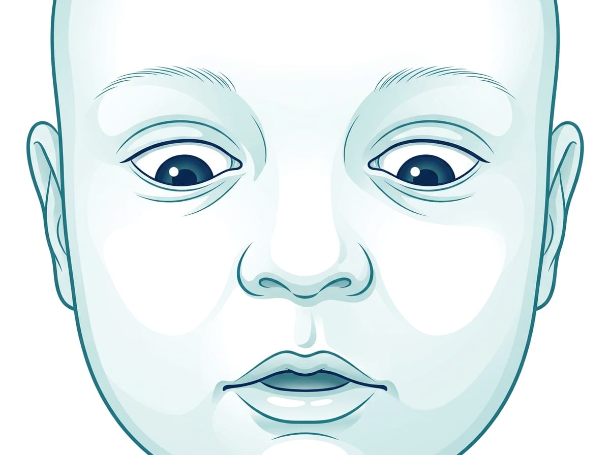
The setting sun sign — sometimes called sunset eyes — is one of the most recognisable visual features of hydrocephalus in infants and young toddlers. The eyes appear pushed downward, with white sclera (the white of the eye) visible above the iris. The upper eyelids may appear retracted.
This sign reflects pressure on the dorsal midbrain — a region controlling upward gaze. When the third ventricle enlarges and presses downward on this structure, upward gaze becomes impaired and the eyes drift into a characteristic downward position.
The setting sun sign is a sign of significant intracranial pressure and requires urgent medical attention when newly observed or when it worsens. In infants with previously treated hydrocephalus, its return can indicate shunt failure.
Pupil Changes and Asymmetry
Unequal pupils — called anisocoria — or pupils that respond sluggishly to light can indicate pressure on the third cranial nerve, which controls pupil constriction. A pupil that is fixed and dilated (blown pupil) in the context of a deteriorating child is a neurosurgical emergency.
Not all pupil asymmetry is dangerous — a small degree of physiological anisocoria is common. What matters is new onset, worsening asymmetry, or asymmetry in the context of other neurological signs. Any parent who notices that their child’s pupil response has changed from a previous baseline should contact their neurology team promptly.
Gaze Palsy and Restricted Eye Movement
Sixth nerve palsy — inability to move the eye outward — is one of the most common eye movement abnormalities in raised intracranial pressure across all ages. Children with sixth nerve palsy may have a noticeable head turn or squint as they compensate for the restricted movement.
Third nerve palsy produces drooping of the upper eyelid (ptosis), a large pupil, and an eye turned downward and outward.
These palsies are caused by pressure on or stretching of the cranial nerves as they travel through the skull. They may resolve when intracranial pressure is treated effectively — but persistent palsy after pressure normalisation warrants specialist ophthalmology assessment.
Nystagmus
Nystagmus — involuntary rhythmic movement of the eyes — can occur in hydrocephalus when the cerebellar or brainstem pathways controlling eye stability are affected. It may be horizontal, vertical, or rotatory depending on which pathways are involved.
In infants, nystagmus can be subtle and is sometimes dismissed as normal developmental variation. In the context of known or suspected hydrocephalus, nystagmus warrants ophthalmological and neurological assessment.
Cortical Visual Impairment
Cortical visual impairment (CVI) occurs when the visual processing areas of the brain — rather than the eyes themselves — are damaged. In hydrocephalus, CVI can result from direct compression of the occipital lobes by enlarged ventricles, or from ischaemia caused by elevated pressure.
Children with CVI may have structurally normal eyes that pass a basic visual assessment, while their visual function in practical terms is significantly impaired. They may have difficulty recognising faces or objects, struggle with complex visual environments, prefer certain colours or movement, and show variable visual function across different settings and times of day.
CVI is frequently missed precisely because the eyes appear normal on examination. Functional vision assessment — observing how a child uses vision in everyday situations — is essential for identifying it. A referral to a specialist in paediatric CVI is appropriate for any child with hydrocephalus who shows unexpected visual difficulties.
Visual Field Defects
Enlarged ventricles pressing on the visual radiations — the pathway carrying visual information from the thalamus to the visual cortex — can produce visual field defects. A child may have good central vision while being unable to see objects in peripheral areas of their visual field.
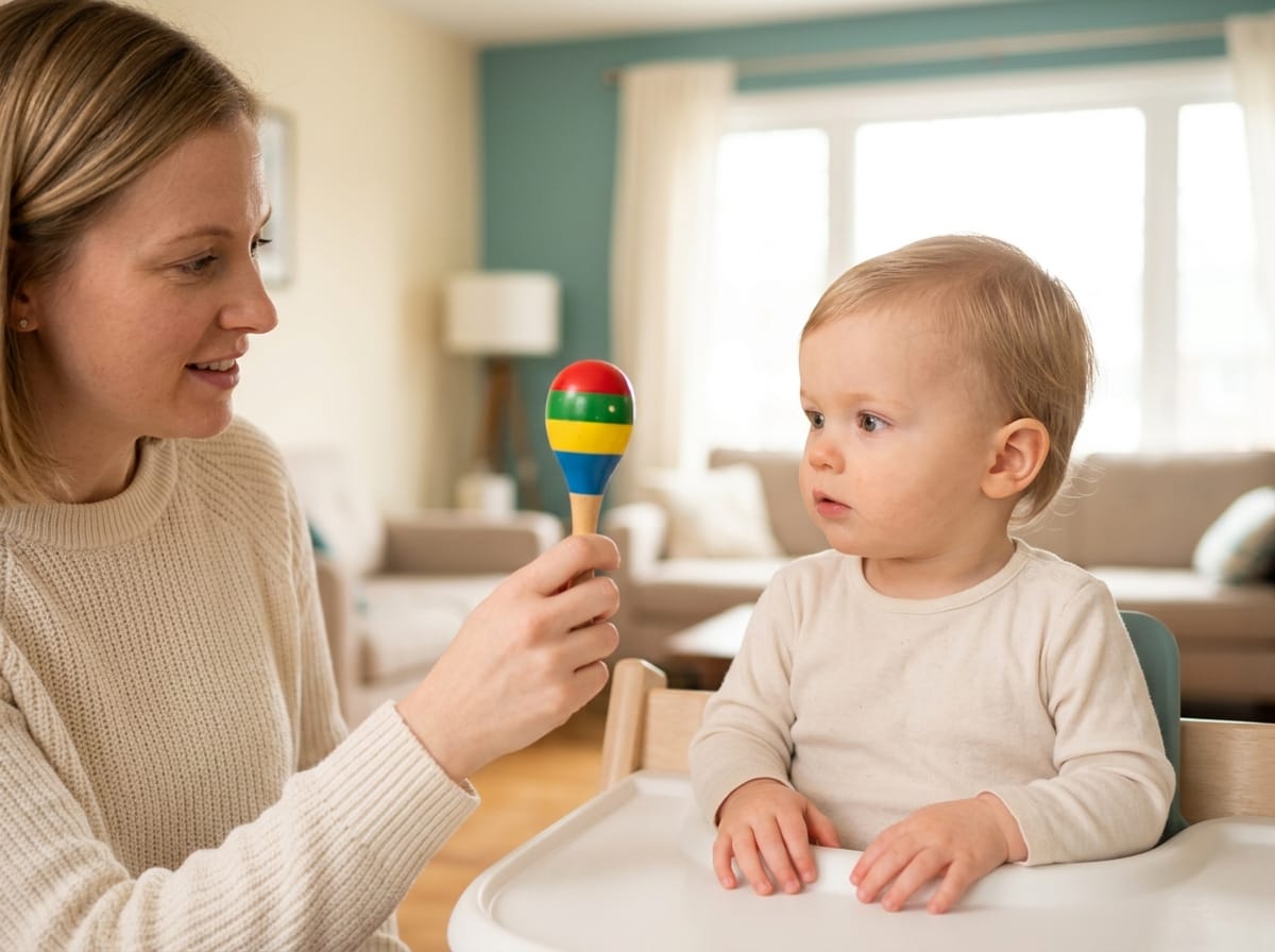
Visual field defects are difficult to identify in young children who cannot cooperate with formal visual field testing. Observation in everyday situations — does the child bump into things on one side, reach only to one side, seem unaware of objects in certain positions — can provide important clues.
What the Research Shows
Research on optic nerve involvement in paediatric hydrocephalus consistently identifies the optic nerve sheath as a pressure transmission pathway that produces measurable changes in optic disc appearance before other signs of raised intracranial pressure become clinically apparent. Studies have demonstrated that optical coherence tomography — a non-invasive imaging technique of the retina — can detect subclinical optic nerve changes in children with hydrocephalus before papilloedema is visible on standard examination. Available at PubMed
A comprehensive review of cortical visual impairment in children identified hydrocephalus as one of the leading causes — along with periventricular leukomalacia and hypoxic-ischaemic injury. The review emphasised that CVI is the most common cause of visual impairment in children in high-income countries and remains significantly under-diagnosed, particularly in children whose structural eye examinations are normal. Available at PubMed
I want to be honest about a limitation: most research on visual outcomes in hydrocephalus comes from studies of congenital or shunt-treated hydrocephalus. Research specifically on visual outcomes in post-infectious hydrocephalus — the type most relevant to our family’s experience — is less comprehensive. The mechanisms described above apply across hydrocephalus types, but the specific trajectory of visual recovery after infection-related pressure may differ from what is described in surgical literature.

What This Means for Your Family
Ask for a baseline ophthalmology assessment at or shortly after your child’s hydrocephalus diagnosis. Not every centre offers this routinely. You may need to ask specifically.
Tell the ophthalmologist the full context — that your child has hydrocephalus, what the ventricular size was, and what treatment has been given. A standard paediatric eye examination without this context may miss pressure-related changes.
At every follow-up neurology appointment, report any visual changes you have noticed at home — even subtle ones. Changes in how your child tracks objects, whether they reach accurately for things, how they navigate familiar spaces, and whether they seem to notice objects at the sides of their vision all provide useful clinical information.
If your child is school-age, share visual concerns with their teacher. A child with unrecognised visual field defect or CVI may appear inattentive or learning-delayed when the primary issue is that they cannot see the parts of the board or page they are expected to be using.
Questions to Ask Your Neurology and Ophthalmology Team
- Has my child had a formal ophthalmology assessment since their hydrocephalus diagnosis?
- Are there signs of papilloedema or optic disc changes on fundoscopy?
- Is there any evidence of cranial nerve palsy affecting eye movement?
- Given my child’s ventricular size and location, which visual pathways are most likely to be affected?
- Should we assess for cortical visual impairment — and if so, who performs this assessment?
- If visual problems are present, will they resolve with pressure treatment, or do they require separate management?
Frequently Asked Questions
Can hydrocephalus cause blindness in children?
Hydrocephalus can cause visual impairment ranging from mild to severe depending on which visual pathways are affected and how long elevated pressure persists. Permanent blindness is uncommon when hydrocephalus is treated promptly. Prolonged untreated elevated pressure can cause lasting optic nerve damage. Cortical visual impairment from damage to visual processing areas can persist even after pressure is normalised.
What is the sunset sign in hydrocephalus?
The setting sun sign is a characteristic eye appearance in infants with raised intracranial pressure — the eyes appear pushed downward with white sclera visible above the iris. It results from pressure on the midbrain structures controlling upward gaze. It is associated with significant intracranial pressure and requires urgent medical attention when newly observed.
Can visual problems from hydrocephalus improve with treatment?
Many visual problems associated with raised intracranial pressure improve when that pressure is effectively treated. Cranial nerve palsies causing squint or restricted eye movement frequently resolve after shunting or other pressure management. Cortical visual impairment may improve with neuroplasticity-driven recovery over months to years, particularly in young children. Some changes, particularly optic nerve damage from prolonged pressure, may be permanent.
Should every child with hydrocephalus see an ophthalmologist?
Yes. A baseline ophthalmology assessment is appropriate for all children with hydrocephalus. The eyes provide direct information about intracranial pressure and optic nerve health that supports neurological monitoring. Specialist assessment for cortical visual impairment is additionally appropriate for any child showing unexpected visual difficulties in everyday function.

The evening I noticed his pupils I called the hospital. Not because I knew precisely what I was looking at — but because something in the pattern of what I had been reading over preceding weeks told me the eyes were connected to the pressure in a way that mattered.
I was right. His pressure had risen. The team adjusted his management that night.
The eyes did not lie. They had been telling us something for days before any other sign was obvious enough to act on.
Learn to read what they are saying. Ask for the assessments that make those signs legible. The visual system is one of the brain’s most direct communication channels — and in hydrocephalus, it is one worth listening to carefully.
This article is for informational purposes only and does not constitute medical advice. Always consult your child’s neurologist and ophthalmologist for assessments specific to your child’s condition. Read our full disclaimer: braincarepath.com/disclaimer/
Medically Reviewed by Dr. Abubakar Siddique, MBBS, FCPS (Neurology) — Consultant Interventional Neurologist, HOD Neurology and Stroke Unit, Akhter Saeed Medical and Dental College, Lahore.
What are hydrocephalus eye symptoms in babies?
In babies, the most recognised sign is the setting sun sign, where the eyes appear driven downward so white is visible above the iris. Other signs include eyes that do not follow faces, unequal pupils, persistent squint after four months, and rapid involuntary eye movement.
Is blurred vision a sign of shunt failure?
It can be. New blurred or double vision in someone with a shunt should be treated as possible shunt failure until proven otherwise, particularly alongside headache, vomiting or drowsiness. Contact your neurosurgical team the same day rather than waiting for a routine appointment.
Bibliography
- Killer HE, Laeng HR, Flammer J, Groscurth P. Architecture of arachnoid trabeculae, pillars, and septa in the subarachnoid space of the human optic nerve. Br J Ophthalmol. 2003;87(6):777-781. Available at PubMed
- Borchert M, Liu GT, Pineles S, Demer JL. Pediatric optic neuritis. J Neuroophthalmol. 2011;31(4):380. Available at PubMed
- Huo R, Burden SK, Hoyt CS, Good WV. Chronic cortical visual impairment in children: aetiology, prognosis, and associated neurological deficits. Br J Ophthalmol. 1999;83(6):670-675. Available at PubMed
- Dutton GN. Cerebral visual impairment: working within and around the limitations of vision. Semin Pediatr Neurol. 2003;10(1):70. Available at PubMed